
Figure 4.
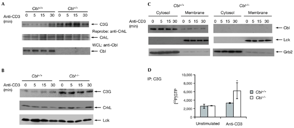
Cbl regulates C3G activation. (A) The CrkL–C3G interaction was increased in Cbl−/− thymocytes. Thymocytes from Cbl+/+ and Cbl−/− mice were either unstimulated or stimulated with anti-CD3 for the durations indicated, followed by immunoprecipitation (IP) with anti-CrkL, blotting, and detection with anti-C3G. The membrane was reprobed with anti-CrkL. (B) Membrane translocation of C3G was increased in Cbl−/− thymocytes. Cells treated as in (A) were subjected to subcellular fractionation and the membrane fraction was immunoblotted with anti-C3G, anti-CrkL or anti-Lck as indicated. (C) The membrane and cytosolic fractions from (B) were immunoblotted with anti-Cbl, anti-Lck or anti-Grb2. (D) Guanine-nucleotide exchange factor assay. Anti-C3G immunoprecipitates from thymocytes treated as in (A) were analysed for exchange activity using recombinant Rap1 as a substrate. The binding of [32P]GTP to Rap1 was measured using a liquid scintillation counter.