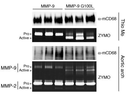
Figure 5.
CD68S vectors drive the overexpression of MMP-9 in macrophages and atherosclerotic lesions in apoE–/– mice. MMP-9 expression in thioglycollate-elicited peritoneal macrophages (Thio) and aortic arch lysates from apoE–/– mice transplanted with HSCs transduced with CD68S–MMP-9 or CD68S–MMP-9 G100L retroviruses was analyzed by gelatin zymography as described in the Figure 2A legend. Immunoblotting with an antibody against macrosialin (α-mCD68) served as loading control for the isolated macrophages and a measure of macrophage content of lesions in the aortic arch. The proforms and active forms of MMP-9 and MMP-2 are highlighted.

