
Figure 2.
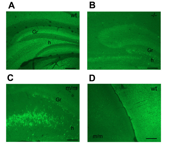
Kv1.1 immunohistochemistry in wild type, Kv1.1-null and mceph/mceph hippocampus. Immunohistochemistry on formalin fixed brain sections. Exposure parameters were adjusted to obtain a strong and clear signal. Note exposure times given to relate between panels A. Wild type hippocampus showed the same staining pattern as that previously reported for Kv1.1 [2] (exposure 1100 ms) B. Kv1.1 null mouse hippocampus: some cross reactivity of the antibody was seen. (exposure 2700 ms) C. In the mceph/mceph hippocampus the immunoreactivity surrounded the nuclei of neurons especially in the dentate gyrus hilus (h) (exposure 2500 ms) D. Parietal neocortex in mceph/mceph (left) and wild type (right) brain: the staining of fibers in wild type was absent in mceph/mceph. Scale bar 200 μm; h, dentate gyrus hilus; Gr, dentate gyrus granular cell layer; wt, wild type; -/-, Kv1.1-null; m/m, mceph/mceph