Figure 2.

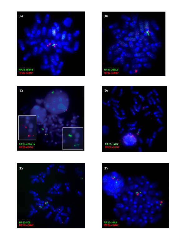
Fluorescence in situ hybridization (FISH) maps the TgAS centromeric and telomeric deletion extent. In each case, BAC RP22-434N7 from the chromosome 7 Tyrosinase (Tyr) locus (74.6 Mb; 44.0 cM) was hybridized as a control and is shown as a red signal. All chromosome 7B/C BACs used as probes are shown as green signals while chromosomes are stained with DAPI (blue). (A) BAC RP24-354P8 spanning the region including Siglec-H and Tubgcp5 shows a single-chromosome 7 signal indicating the deletion of this locus in TgAS splenocytes. (B) RP23-256L9 spans the p locus (exons 10–24) and is also deleted in TgAS mice. (C) RP24-426A19 spans the region 3' of Luzp2 and detects a weak signal on one chromosome 7 homologue, suggesting partial deletion and detection of the centromeric breakpoint in the mutant mice. The two insets show the images for individual probes. (D) RP23-199N11 covers part of the region between Frat3 and Chrna7 and is deleted in TgAS mice. (E) RP23-506 for the Klf13 locus is intact in the deletion mice. (F) RP23-16A4 spans all 10 exons of Chrna7 and shows an apparently intact signal in TgAS mice.
