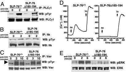
Fig. 6.
Phosphorylation of PLC-γ1, Itk, Vav, and Erk, and calcium flux after TCR/CD3 ligation in splenic T cells. Cells were stimulated with anti-CD3 (5 μg/ml), washed, and crosslinked with 10 μg/ml goat F(Ab′)2 anti-rat Ig. Lysates were immunoprecipitated with anti-PLC-γ1 (A), anti-Itk (B), or anti-Vav1 (C) Abs, run on 10% SDS/PAGE gels, probed with antiphosphotyrosine (pTyr) mAb 4G10, and reprobed with anti-PLC-γ1, anti-Itk, or anti-Vav Abs as loading controls. (D)Ca2+ mobilization: Purified T cells preloaded with fura-2 acetoxymethyl ester were stimulated with biotinylated anti-CD3 and crosslinked with streptavidin (SA) then later stimulated with ionomycin (io) in 2 mM Ca2+ Ringer's solution. (E) Erk phosphorylation. Lysates from purified spleen T cells stimulated with crosslinked anti-CD3 as described in A were probed with phospho-ERK-specific Ab (pErk) and reprobed with Erk-specific Ab. The results shown are representative of three experiments for A–C and two experiments for D.

