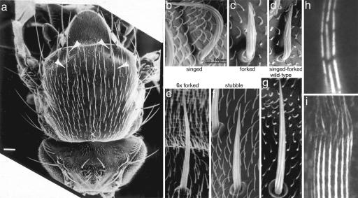
Fig. 1.
Scanning electron and light micrographs of fly bristles and hairs. (a) The thorax of an adult, wild-type fly. The arrows point to four macrochaete bristles. The shorter bristles are microchaetes. (Scale bar, 100 μm.) [Reprinted with permission from ref. 6 (Copyright 1995, The Rockefeller University Press).] (b–g) Representative microchaetes from mutant flies. Surrounding the microchaetes are the much shorter hairs. [Reprinted with permission from ref. 3 (Copyright 2004, American Society for Cell Biology).] (h) Light micrograph of fluorescent bundles from wild-type, 48-h-old pupae. Note that the bundles are made of modules, which tend to be in transverse register. [Reprinted with permission from ref. 4 (Copyright 1996, The Rockefeller University Press).] (i) Light micrograph from bundles in a bristle from a 33-h-old pupa. The bundles are stained with fluorescent antibody against the forked protein. Note how the gaps in the modules fill in as one moves from the tip to the base. (Scale bar, 5 μm.) [Reprinted with permission from ref. 11 (Copyright 2003, The Rockefeller University Press).]

