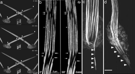
Fig. 3.
Images of bristles taken with fluorescently labeled actin. (a) Images taken at three different times after stripes are bleached in the bristles. Note that the stripes move toward the base. Insets show that there is recovery of fluorescence in the bleached area (courtesy of G. M. Guild). (b) Two time-lapse images taken of the same disassembling bundles. Note that disassembly takes place from the ends of filaments nearest the tip, which are the barbed ends. (Reprinted from ref. 25). (c and d) Images of a bundles treated from cells treated with jasplakinolide. Note that the bundles are translocated in from the bristle into the cytoplasm. [Reprinted with permission from ref. 16 (Copyright 2003, American Society for Cell Biology).]

