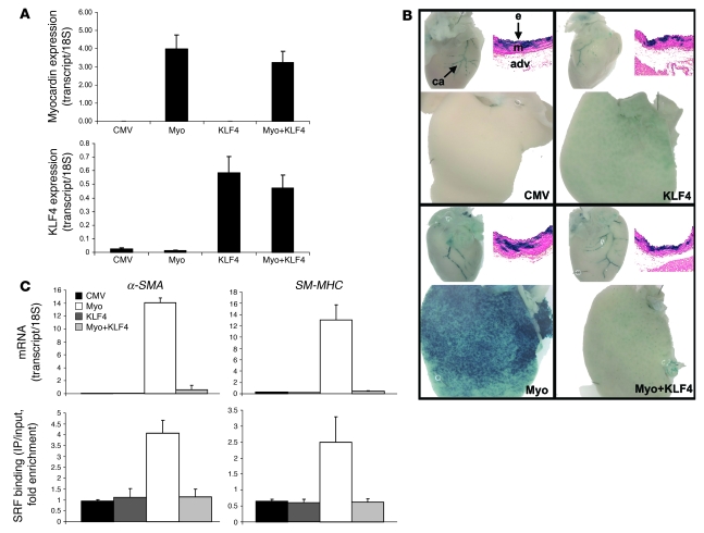
Figure 5. Myocardin and KLF4 exert opposing influences over SMC gene expression in transgenic mouse liver in vivo.
(A) mRNA was extracted from liver in mice infected with CMV-empty control viruses, CMV-KLF4, CMV-myocardin, or mice coinjected with myocardin and KLF4 viruses. Expression levels of myocardin and KLF4 were measured by real-time PCR to document that delivery of these genes was successful. Data were normalized to levels of 18S expression. (B) LacZ staining from SM-MHC-LacZ–transgenic mice injected as in A. For each panel in B, the top left image shows hearts expressing SMC-specific LacZ staining in coronary arteries (ca), and the top right image shows cross-sections taken from these mice displaying SMC-specific LacZ staining in the media (m) of aortas. The endothelial layer (e) and adventitia (adv) are labeled. The media exhibit mosaic staining, which is typical for SM-MHC–transgenic mice. The bottom image in each panel shows staining for the presence of LacZ in mouse liver. (C) mRNA levels of α-SMA and SM-MHC were measured by real-time RT-PCR, and SRF binding to CArG box chromatin of these genes was measured by ChIP, in livers of mice injected with the indicated corresponding adenoviruses.

