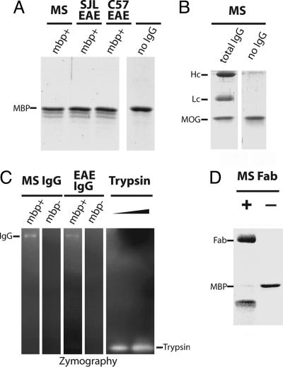
Fig. 2.
MBP degradation by purified autoantibodies. MBP affinity-purified antibodies from sera of the MS patient and EAE SJL and EAE C57BL/6 mice were incubated with purified MBP (A) and MOG (B) followed by SDS/PAGE analysis and Coomassie staining. IgG mbp+, MBP-binding antibodies. IgG heavy chains, light chains, and noncleaved MBP bands are marked as Hc, Lc, and MBP, respectively. (C) The MBP autoantibodies were separated by 5-20% gradient SDS/PAGE with FITC-BSA fluorescent substrate impregnated in the separating gel. After electrophoresis, proteins were in-gel renatured by Triton X-100 washes. Proteolytic degradation was visualized by fluorescence intensity increase, seen as bright bands on the dark background. Bovine trypsin (10 and 30 pg) was used as a positive control. IgG mbp+, MBP-binding antibodies; mbp-, antibodies not bound to the MBP-affinity column. (D) MBP degradation by the purified Fab fragment derived from the whole IgG of the MS patient visualized by SDS/PAGE.

