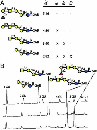
Fig. 7.
Globo H structural confirmation by analytical sequence analysis. (A) Shown are the glycans obtained by exoglycosidase cleavage with the indicated enzymes (E1 = α-fucosidase, E2 = β1,3-galactosidase, E3 = β-N-acetylhexosaminidase), marked by an X, and the glucose unit (GU) value relative to fluorescently labeled dextran standard. (B) Sample chromatograms from normal phase-HPLC with fluorescence detection (excitation = 330 nm, emission = 420 nm) highlighting glycans obtained during sequence analysis.

