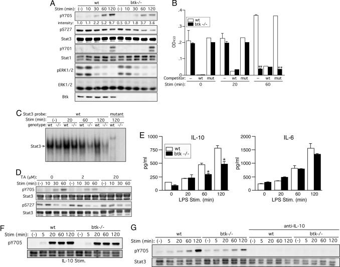
Fig. 6.
Btk positively regulates Stat3 activity mainly through autocrine secretion of IL-10. (A) WT and btk–/– BMDCs were left unstimulated or stimulated with 1 μg/ml LPS for the indicated periods. The cells were lysed and analyzed by immunoblotting with anti-phospho-Stat3(Tyr-705), anti-phosho-Stat3(Ser-727), anti-phospho-Stat1(Tyr-701), anti-phospho-MAPK(Thr-202/Tyr-204), or anti-Btk. The blots were reprobed with anti-Stat3, Stat1, and anti-ERK to check for their expression. Results shown are representative of seven (Stat3) or three (others) experiments. (B) DNA-binding activities of Stat3 in BMDCs stimulated by LPS for the indicated periods were measured by ELISA-based binding assays. Specificity of Stat3 binding to the plate-bound Stat3 consensus oligonucleotide was confirmed by strong competition by free WT, but not mutant, oligonucleotide. **, P < 0.005 (vs. the WT cells; Student's t test). Similar results (mean ± SD) are reproduced in three independent experiments. (C) EMSAs were also performed by using 32P-labeled WT or mutant oligonucleotide as a probe. Position of Stat3/DNA complexes is indicated. Similar results are reproduced in three independent experiments. (D) WT BMDCs were preincubated with the indicated concentrations of terreic acid (TA) for 30 min before stimulation with LPS for the indicated periods. The cells were analyzed by immunoblotting with anti-phospho-Stat3(Tyr-705) or anti-phospho-Stat3(Ser-727). The blots were reprobed with anti-Stat3. The results shown were reproduced in two additional experiments. (E) Wt and btk–/– BMDCs were left unstimulated or stimulated with 100 ng/ml LPS for the indicated periods. Amounts of IL-10 and IL-6 secreted into medium were measured by ELISA. *, P < 0.05 (vs. the WT cells; Student's t test). Similar results (mean ± SEM) were reproduced in another experiment. (F) WT and btk–/– BMDCs were left unstimulated or stimulated with 20 ng/ml mouse recombinant IL-10 for the indicated periods. (G) WT and btk–/– BMDCs, pretreated with a neutralizing anti-IL-10 mAb for 60 min, were left unstimulated or stimulated with 1 μg/ml LPS for the indicated periods. Cell lysates were analyzed by immunoblotting for Tyr-705 phosphorylation followed by reprobing with anti-Stat3. Results shown in F and G are representative of two independent experiments.

