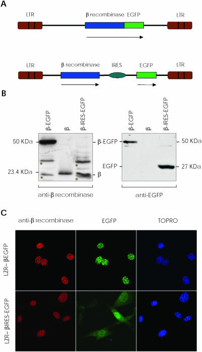
Figure 1.
β Recombinase expression using retroviral vectors. (A) Scheme of LZR-β-EGFP and LZR-β-ires-EGFP retroviral vectors used. Expression of a direct fusion protein between β recombinase and EGFP (upper); independent expression of both proteins using an IRES (lower). Arrows indicate the transcribed sequence. (B) NIH-3T3 cells were transduced with both vectors; after 48 h, total proteins were extracted. Western blot autoradiograph shows expression of the fusion protein (β-EGFP, 50 kDa, left lane), individual proteins (β, 23.4 kDa; EGFP, 27 kDa, right) and 5 ng of purified β recombinase (center). Non-specific bands are marked with an asterisk on the membranes. (C) Immunofluorescence of NIH-3T3 cells transduced with LZR-β-EGFP (upper) and LZR-β-ires-EGFP (lower), showing nuclear localization of β recombinase, fused or alone. EGFP is detected in the same nuclear dots when LZR-β-EGFP is used, but not when β recombinase and EGFP are expressed independently (LZR-β-ires-EGFP). TOPRO stains nuclei.

