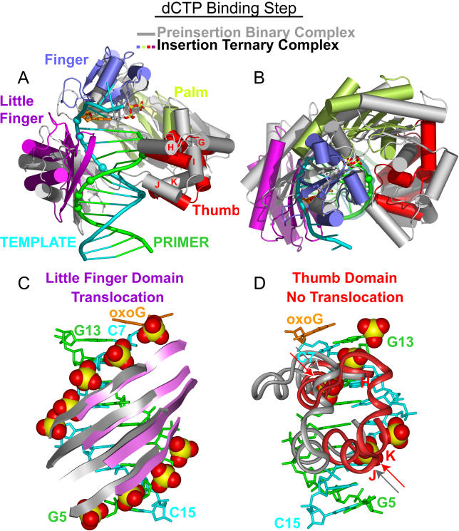
Figure 4. Conformational Transitions of Dpo4 and oxoG-Modified Template-Primer DNA Associated with the Nucleotide-Binding Step Following Superimposition of DNA Duplexes.
(A) Overall comparative views normal to the DNA helix axis (following superimposition of the DNA duplexes) of the structures of the preinsertion binary complex (in silver) and the insertion ternary complex with incoming dCTP (in color). The DNA-backbone phosphate groups in contact with the Dpo4 little-finger and thumb domains in the ternary complex are shown by colored spheres.
(B) Overall comparative view looking down the DNA helix axis.
(C) Comparative views of contacts between the little-finger domain and the DNA backbone, with contacts shown by CPK spheres. The β-sheets of the little-finger domain move by a one nucleotide step upon proceeding from preinsertion binary complex (silver) to insertion ternary complex (purple).
(D) Comparative views of contacts between the thumb domain and the DNA backbone, with contacts shown by CPK spheres. The α-helical segments of the thumb domain (helixes H, K, J) retain the same contacts with the backbone phosphates of the primer and template strands (shown by arrows) upon proceeding from preinsertion binary complex (silver) to insertion ternary complex (red).

