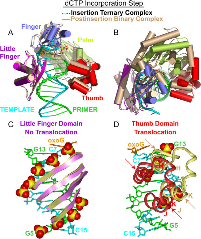
Figure 7. Conformational Transitions of Dpo4 and oxoG-Modified Template-Primer DNA Associated with the Covalent Incorporation Step Following Superimposition of DNA Duplexes.
(A) Overall comparative views (following superimposition of DNA duplexes) of the structures of the insertion ternary complex with incoming dCTP (in color) with the postinsertion binary complex after covalent cytosine incorporation (in beige). The DNA-backbone phosphate groups in contact with the Dpo4 little-finger and thumb domains in the ternary complex are shown by colored spheres.
(B) Overall comparative view looking down the DNA helix axis.
(C) Comparative views of contacts between the little-finger domain and the DNA backbone are maintained upon proceeding from the insertion ternary complex (purple) to the postinsertion binary complex (beige).
(D) Comparative views of contacts between the thumb domain and the DNA backbone, which shift by a one nucleotide step (shown by arrows) upon proceeding from the insertion ternary complex (purple) to the postinsertion binary complex (beige).

