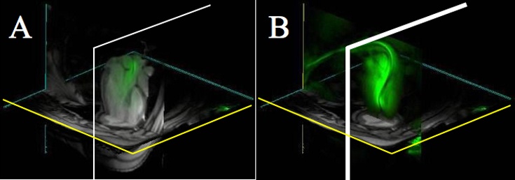
Figure 2.

Catheter tracking using 3D multislice MR fluoroscopy: A, Thin slice of catheter (green) within LV. Distal end is outside of scan plane and therefore not visible. B, By switching to a catheter-only mode, entire catheter becomes visible, analogous to x-ray projection.
