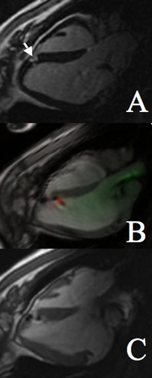
Figure 4.

Precise targeting of infarct border of a small infarct. A, Conventional inversion recovery DHE showing small area of infarction (arrow). B, Real-time steady-state free precession image of second injection into infarct border. C, Postinjection high-resolution gated, breath-held steady-state free precession image clearly showing 2 border injections.
