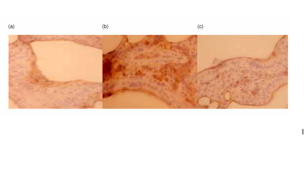
Figure 6.

Immunohistochemical staining with TGF-β antibody on murine lung tissues. Three mice from each group were checked. Representative slides are shown for (a) unirradiated control mice at 3 weeks after sham-irradiation; (b) untreated mice at 3 weeks after 10 Gy irradiation; (c) CAPE-treated mice at 3 week after irradiation 10 Gy irradiation. These demonstrated that CAPE treatment attenuated the increased TGF-β immunoreactivity after irradiation. Magnification × 250.
