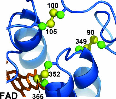
Fig. 1.
Structure of the Ero1p active site. A ribbon diagram of the active-site region of yeast Ero1p (4) is shown with the bound FAD in orange and disulfides in a ball-and-stick representation. The Cys sulfurs are numbered according to the position of the residue in the yeast Ero1p sequence (RefSeq accession no. NP_013576). The disulfide between Cys-352 and Cys-355 abuts the flavin. The loop containing Cys-100 and Cys-105 is flexible (only one of the two conformations observed in the crystal structures is shown) and may be involved in shuttling electrons from substrate to the Cys-352-Cys-355 disulfide. The structural or functional role of the Cys-90-Cys-349 disulfide has yet to be determined.

