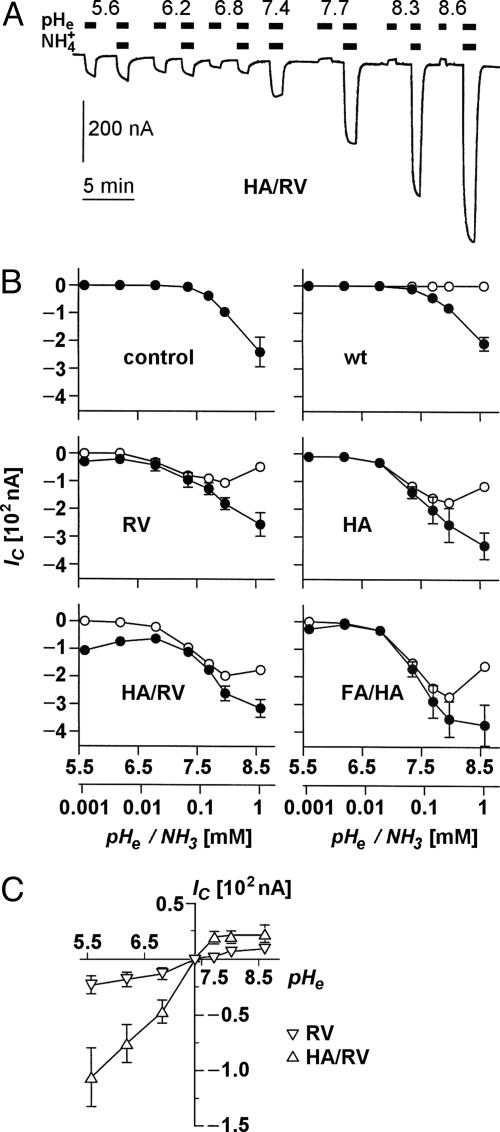
Fig. 4.
Ammonia- and pH-induced proton currents in Xenopus oocytes expressing AQP1 wild-type and mutants. (A) Sample trace of clamp current (-50 mV) induced in an AQP1-H180A/R195V-expressing oocyte by changes in pHe and  of the bathing solution. The baseline current was -100 nA. pHe was intermittently changed from 7.4 (control) to a range of values from 5.6 to 8.6 ± a concurrent change in
  (5 mM Na+ was replaced by 5 mM
  ), as indicated by the bars on top. In the acidic range, pHe changes resulted in inward currents that were independent of the presence of
  . In the alkaline range, changes in pHe resulted in small outward currents, whereas simultaneous addition of
  induced large inward currents. The LP of the oocyte was 9.6·10-3 cm·s-1. (B) Filled symbols: total clamp currents (-50 mV) induced by 5 mM
  at various pHe values in uninjected oocytes (control) and oocytes expressing AQP1 wild-type and mutants. The open symbols represent the currents induced by AQP-facilitated ammonia passage, i.e., we subtracted the background current induced by ammonia diffusion (control, Top Left) and the current produced by ammonia-independent proton permeation of certain AQP1 mutants (depicted in C). (C) Clamp currents elicited exclusively by changes in pHe. For clarity, we show only the currents for AQP1-R195V and AQP1-H180A/R195V. The currents for AQP1 wild-type and the AQP1-H180A mutant were not significantly different from the miniscule currents already observed in uninjected control oocytes. The current for AQP1-F56A/H180A (not shown) was only significantly different at pHe 5.6, i.e., 17 ± 2 nA (n > 5 for AQPs and n = 3 for uninjected controls).

