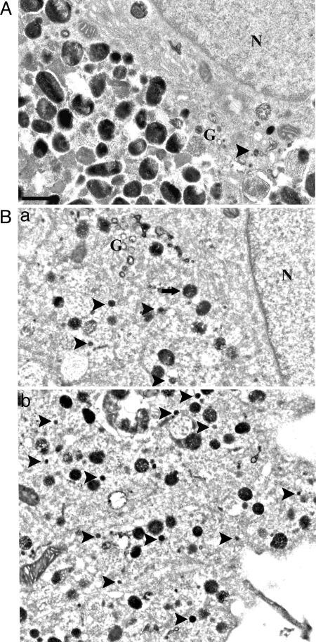
Fig. 4.
Ultrastructural analysis of positive DOPA reaction products. (A) DOPA histochemistry identified Golgi tubules (G) in control melanocytes. The 50-nm vesicles were infrequent and confined to the Golgi area. (B) In DAPT-treated melanocytes, numerous 50-nm vesicles (arrowheads) could be identified close to Golgi area (G), throughout the cell body (a), as well as in dendrites (b). (Scale bar: 0.5 μm.)

