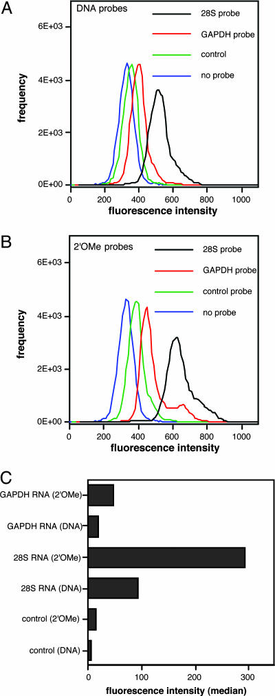
Fig. 3.
Detection of GAPDH mRNA and 28S rRNA in living HL-60 cells by FC. QFRET probe sequences are shown. The control probe pair consisted of donor probe for 28S rRNA and acceptor probe for GAPDH. HL-60 cells permeabilized by SLO were incubated with QFRET probes (200 nM) in PBS buffer (pH 7.0) for 1.5 h. The resulting cell suspension (n = 50,000) was directly analyzed by FC as described in Materials and Methods.(A and B) Histograms showing cell-count frequency vs. FRET intensity for each probe sequence. (C) Medians of FRET intensity calculated from the histograms. The data were corrected by cell autofluorescence background values (no probes).

