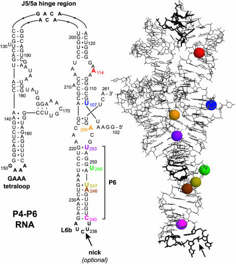
Figure 2.
Secondary and tertiary structure of P4–P6 (22). Colored spheres highlight 2′-hydroxyls of the eight pyrene-labeled nucleotides that are marked on the secondary structure. The backbone may be nicked within the L6b loop without perturbing the tertiary folding (25), which permits assembly of full-length P4–P6 without covalent ligation of the 136-nt and 24-nt fragments (Figure 3).

