Figure 4.

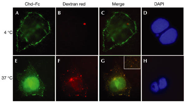
Chordin–Fc translocates into intracellular endocytic compartments at 37 °C. Cells were incubated on ice with Chordin (Chd)–Fc for 2 h, washed, and then incubated with Texas-Red–dextran for 30 min on ice (A–D) or at 37 °C (E–H). (A,E) Localization of Chd–Fc (green) bound to COS-7 cells. (B,F) Localization of Texas-Red–dextran (red) in punctate endocytic compartments after shifting to 37 °C. (C,G) Merged images. (D,H) 4,6-diamidino-2-phenylindole (DAPI) staining of nuclear DNA. Note that at 4 °C, most of the Chd–Fc is detected at the cell surface, but after 30 min at 37 °C it colocalizes with endocytic vesicles in the cytoplasm that contain Texas-Red–dextran (G). The red spot in (B) is artefactual, but shows that red fluorescence does not bleed into the green fluorescence channel.
