
Figure 4.
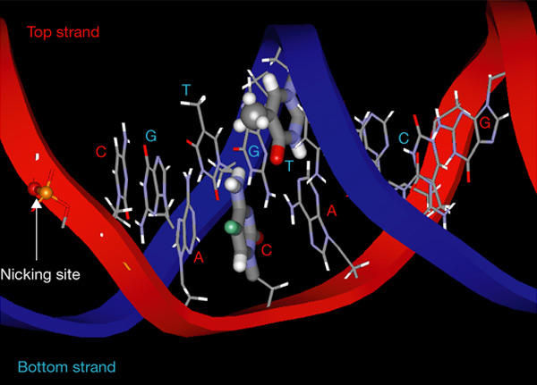
Three-dimensional structure of the consensus heptamer sequence CACAGTG. The bases of the heptamer region are shown by a stick model. The phosphodiester bond where RAG1/RAG2 introduces a nick is indicated. The third cytosine of the top strand and the fourth thymidine of the bottom strand are shown by thick lines. The site of methylation on the third cytosine is shown in green. The methyl group of the thymidine base on the bottom strand is shown as a ball model.