
Figure 3.
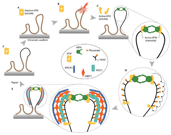
Model of the double-stranded-break response cycle. (A) Undamaged section of a chromosome, showing two chromatin loops and an inactive ATM dimer. (B,C) Induction of a DNA double-stranded break (DSB), modification of chromatin, activation of ATM and recruitment of both ATM and MRE11/RAD50/NBS1 (MRN). The possibility that MRN binds before ATM is shown, but the exact order of events is unknown. The thin black line indicates modified chromatin. (D,E) A wave of H2AX phosphorylation is followed by recruitment of mediators (mediator of DNA damage checkpoint protein 1 (MDC1), p53-binding protein 1 (53BP1) and breast-cancer-associated protein 1 (BRCA1)) to the growing focus, and their ATM-dependent phosphorylation. The molecular architecture of the focus is unknown. (F) Disassembly of the focus, ATM inactivation and chromatin remodelling. The model suggests at least two distinct forms of soluble ATM: an inactive oligomer and an active monomer, and at least two distinct active, insoluble forms: one directly at the lesion and another integral to the growing focus. Note that the MRN complex is also a component of the growing focus but, for clarity, has been omitted here. Complex, persistent lesions are thought to be more difficult to repair, and this is reflected in the size attained by the growing focus. See text for further details. The inset contains a colour-coded key to the molecules shown.