
Figure 3.
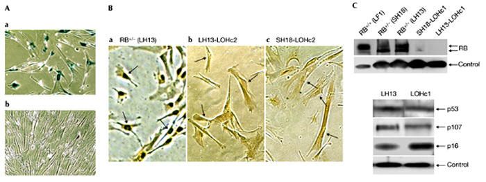
Loss of heterozygosity in RB+/− human fibroblasts. (A) Photomicrographs taken at the time when colonies began to appear. (a) Senescent monolayer of RB+/− cells; (b) a colony with proliferating cells. Both images were taken from the same plate that had been stained for senescence-associated β-galactosidase activity. (B) In situ immunohistochemical detection of RB protein. Colonies of proliferating cells were removed from the primary plates with cloning rings, placed in 24 microtitre wells and after several days of culture expanded into 6-cm dishes. Cells were processed for in situ immunodetection of RB protein after the cultures became established. (a) Presenescent, proliferating LH13 (RB+/−) cells are shown as a positive control. (b) LH13-LOHc2 is a derivative of LH13, and (c) SH18-LOHc2 is a derivative of SH18. Arrowheads point to representative nuclei. (C) Immunoblot analysis. Upper panel, colonies of proliferative cells were picked and expanded as in (B) and immunoblotted with an antibody to RB protein. Cell-line designations are indicated above the lanes. LH13-LOHc1 is a derivative of LH13, and SH18-LOHc1 is a derivative of SH18. Lower panel, expression of p53, p107 and p16 before and after RB LOH. Control, actin; LOH, loss of heterozygosity; RB, retinoblastoma.