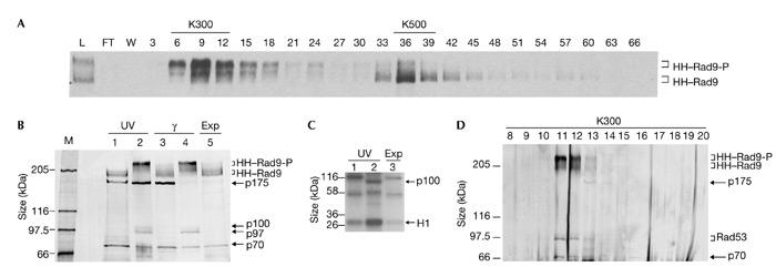
Figure 2.

Purification of HH–Rad9 from untreated and DNA-damaged exponentially growing cells. (A) Rad9 western blot of fractions from a heparin–sepharose column loaded with clarified crude extracts from γ-irradiated cells (750 g). Every third fraction is shown. The 300 mM (K300) and 500 mM (K500) KCl pools and hyperphosphorylated (HH–Rad9-P) and hypophosphorylated (HH–Rad9) epitope-tagged Rad9 are indicated. (B) Silver-stained 6.5% SDS–polyacrylamide gel of the final Ni2+–NTA–agarose purification step. Rad9 and co-purifying polypeptides from cells irradiated with ultraviolet light (UV), γ-irradiated cells and exponentially growing (Exp) cells are indicated. The material loaded in lanes 1 and 3 was derived from K500 heparin–sepharose pools, which predominantly contained hypophosphoylated Rad9, and that in lanes 2 and 4 was derived from K300 heparin–sepharose pools, which predominantly contained hyperphosphoylated Rad9. The relative migration positions of co-purifying 175-, 100-, 97- and 70-kDa polypeptides are indicated. (C) Kinase assay using the purified material from cells irradiated with ultraviolet light and from exponentially growing cells. Lanes 1 and 2 contain material purified from the K500 and K300 heparin–sepharose pools, respectively. The migration position of exogenously added histone H1 and a 100-kDa band (p100) are indicated. (D) Silver staining of fractions obtained after Superose 6 (Pharmacia) gel-filtration of the purified Rad9 complex shown in lane 4 in (B). FT, flow-through; L, load; M, marker; W, wash with lysis buffer.
