
Figure 3.
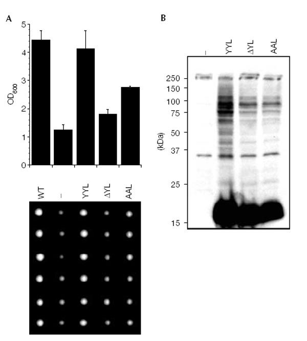
C-terminal HUB1 mutants. (A) The growth of WT Σ1285b cells was compared to hub1Δ (Σ1285b) cells transformed with an empty plasmid (−) or plasmids coding for 3HAHUB1 (YYL), or mutants 3HAHUB1ΔYL (ΔYL) and 3HAHUB1AAL (AAL) under control of the HUB1 promoter. Cells were grown to saturation and then diluted in fresh YPD medium. After 24 h of growth at 30°C, the optical densities were determined at 600 nm (upper panel). Alternatively, single cells from saturated cultures were placed on an agar plate using a micromanipulator and grown at 30 °C until colonies became visible (lower panel). (B) Formation of HUB1–protein adducts with the HUB1 variants described in (A).