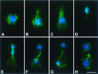
FIG. 1.
Microtubule cytoskeleton and nuclear position during the C. neoformans cell cycle. Immunofluorescent imaging of microtubules (anti-α-tubulin antibodies) was merged with nuclear staining (DAPI). Individual cells are arranged in their presumed order during the cell cycle (from panel A to panel H). Bar, 5 μm.

