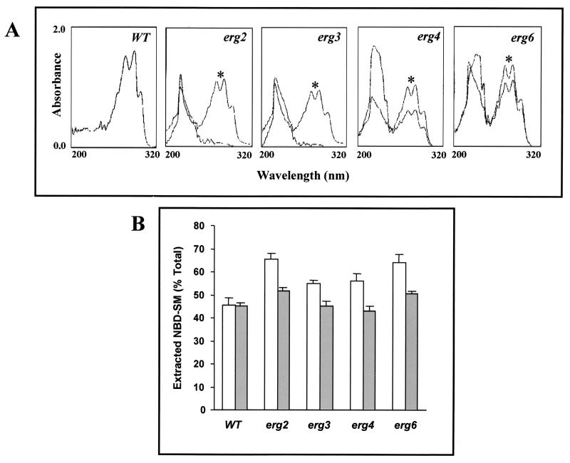
FIG. 7.
(A) UV absorption spectra of sterols extracted from the S. cerevisiae WT strain and its erg mutants grown in the absence and in the presence (∗) of ergosterol. The cells were supplemented with 10 μg of ergosterol ml−1 and grown for 20 h at 30°C before lipid extraction. The supplemented cells were harvested and washed thoroughly before extraction of sterols as described in Materials and Methods. The UV spectra of WT cells were the same as those of the unsupplemented cells; hence, only one trace is shown in the first panel. (B) Postlabeling transbilayer exchange of NBD-SM in the S. cerevisiae WT and its erg mutants. Cells were grown in the absence (open bar) and presence (filled bar) of ergosterol-supplemented media as described in Materials and Methods. NBD-SM labeled cells were washed twice with buffer A and were then incubated with 2% BSA to back exchange the NBD-SM from the labeled cells, as described in Materials and Methods. The percentage of total extracted NBD-SM was calculated (according to the formula given in Materials and Methods). The graph presents data for the 90-min time point during which the maximum back-exchanged fluorescence in the supernatant was observed. The values are the means ± standard deviations (indicated by the bars) of three independent experiments.

