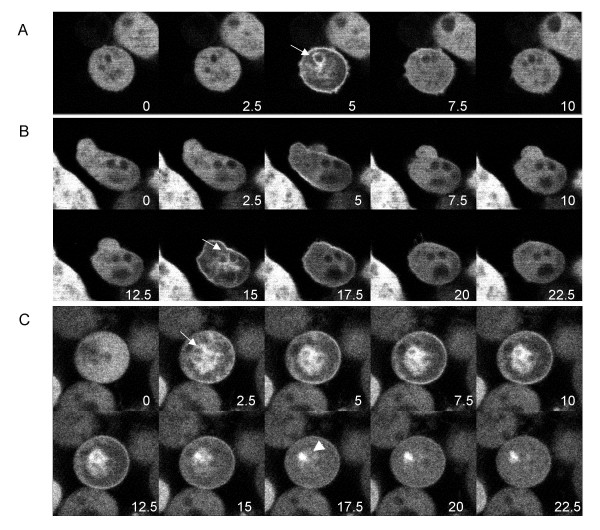
Figure 4.
GFP-CpnA transiently binds to the plasma membrane and intracellular vacuoles in a small subset of starved cells. Cells expressing GFP-CpnA were washed three times in starvation buffer, placed on glass bottom plates, and imaged using a confocal microscope. A) and B) Successive time-lapse images taken every 2.5 seconds of a single cell from a plate of cells starved for 5.5 hours (arrows point to intracellular vacuoles). C) Successive time-lapse images taken every 2.5 seconds of a single cell from a plate of cells starved for 9.5 hours (arrowhead points to fluorescent dot). See Additional Files – Movies 1, 2, 3, 4, 5.

