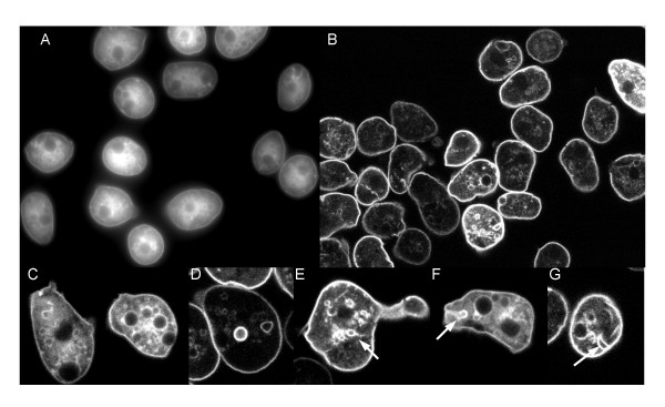
Figure 6.
GFP-CpnA associates with the plasma membrane and intracellular membranous vacuoles in fixed cells. Cells expressing GFP-CpnA were flattened with agarose, fixed, and imaged using A) widefield fluorescence and B) confocal microscopy. Confocal images of single cells are shown in C, D, E, F, and G. Arrows point to labeled vacuoles with attachments to the plasma membrane. G) see Additional File – Movie 9 of z-series images.

