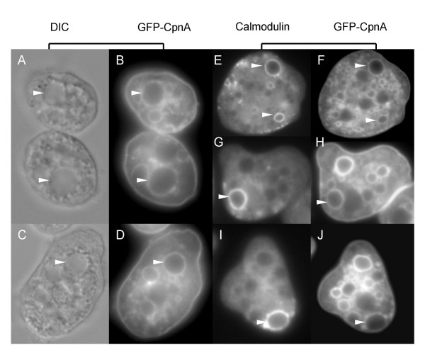
Figure 7.
GFP-CpnA labels contractile vacuoles. Cells expressing GFP-CpnA were placed in water for 1.5 minutes, fixed, and imaged using differential interference contrast microscopy (A, C). These same cells were imaged for GFP with a widefield fluorescence microscope (B, D). Contractile vacuoles in cells expressing GFP-CpnA were labeled with a primary antibody to calmodulin and a TRITC-conjugated secondary antibody and then imaged for TRITC (E, G, I). These same cells were imaged for GFP (F, H, J). Arrowheads point to contractile vacuoles.

