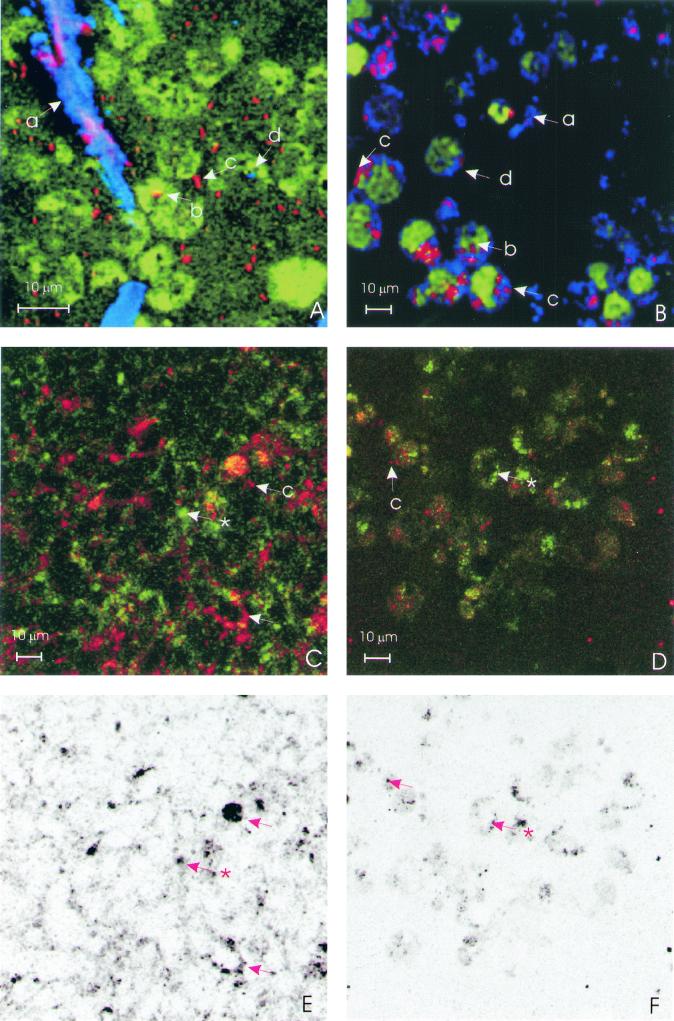
FIG. 2.
Confocal laser scanning microscopy images (all images at ×800 magnification). Bacteria are always red. (A) SV-HUC-1 cell line stained with viable stain Syto-16 with reflecting crystals (blue or purple). (B) HT29-MTX stained with viable stain Syto-16 (green), with reflecting crystals (blue). The letters a, b, c, and d refer to extracellular crystals, intracellular bacteria, extracellular bacteria, and intracellular crystals, respectively. (C) HT29-MTX cell line showing HCM in green and colocalization in yellow (✽, HCM along the cellular membrane; arrow, bacterial and HCM colocalization). (D) HT29-MTX cell line showing HGM in green and colocalization in yellow (✽, HGM without bacterial colocalization; arrow, HGM with bacterial colocalization). The yellow colocalization signal is produced by a simultaneous red TRITC and green FITC signal. (E) Colocalization signal of P. mirabilis and HCM of panel C indicated in black. (F) Colocalization signal of P. mirabilis and HGM of panel D indicated in black.

