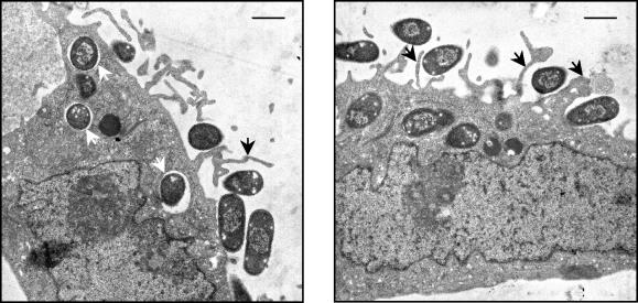
FIG. 4.
Electron micrographs of InvX-expressing E. coli invading HeLa cells. E. coli cells expressing InvX were incubated with HeLa cells for 3 h at an MOI of 10:1 before they were processed for transmission electron microscopy. The micrographs show that E. coli cells attached to the HeLa cell surface elicit membrane protrusions (black arrows) and that intracellular bacilli are located within membrane-bound compartments (white arrows). Scale bars are 1 μm.

