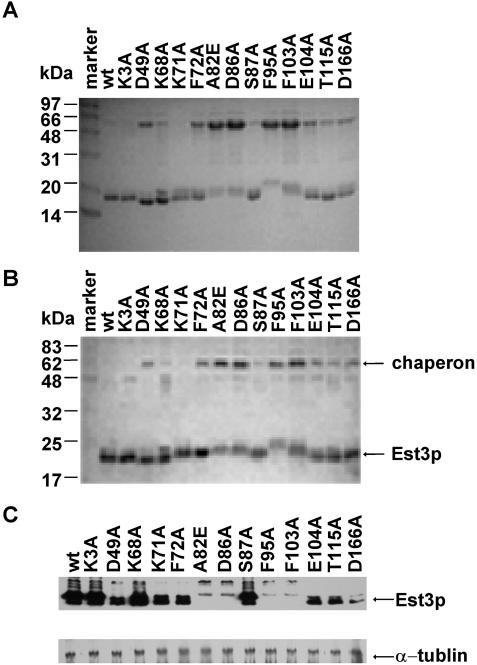
Figure 4.
Purification of wild-type and mutant Est3 proteins. The purified proteins were analyzed with SDS–PAGE stained by (A) Coomassie blue and (B) western blot using affinity purified polyclonal anti-Est3p antibody. (C) The over-expression of Est3 mutants in S.cerevisiae. The upper panel is a western blot probed with affinity purified polyclonal anti-Est3p antibody. The lower panel shows a western blot of the α-tubulin in the same membrane as used in the upper panel (loading control) probed with a monoclonal anti-α-tubulin antibody.

