
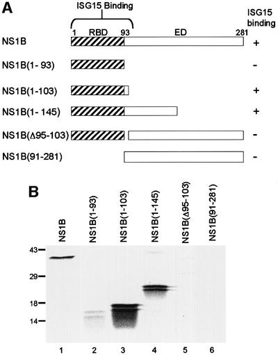
Fig. 2. Identification of the ISG15 binding site on the NS1B protein. (A) Schematic diagram of the fragments of the NS1B protein that were used in the binding assay. RBD, RNA-binding domain; ED, effector domain. (B) Binding assay. GST–ISG15 protein (1 µg) was incubated with 104 c.p.m. of 35S-labeled full-length NS1B protein (lane 1), or the indicated fragment of the NS1B protein (lanes 2–6) in the presence of 20 µl of glutathione–Sepharose 4B beads. The same amount of each radiolabeled NS1B fragment was added to the binding assay. The labeled proteins eluted from the beads were analyzed by electrophoresis on a 12% SDS–polyacrylamide gel. The doublets seen with the NS1B fragments (lanes 2–4) were present both before and after the binding assay. The mobilities of molecular weight markers are shown on the left.