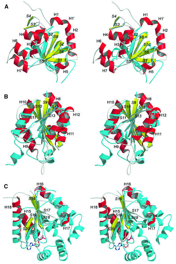
Fig. 3. Stereoviews of ATPS domains I, II and IV (helices, red; strands, yellow; coiled regions or turns, grey) in ribbon presentation showing a structural comparison of the typical fold motifs of the single domains with the best-fitting members of the related superfamilies (cyan). (A) Domain I superimposed with the pyruvate kinase β-barrel domain. (B) Superimposition of domain II with GCT, a typical member of the superfamily of nucleotidylyl transferases. Both polypeptides show the typical α/β-fold of the nucleotidylyl transferases. (C) Overlay of the structure of domain IV with yeast uridylate kinase, complexed with ADP. Both polypeptides show the typical nucleotide and nucleoside kinase fold. All ATPS secondary structure elements are labelled.
