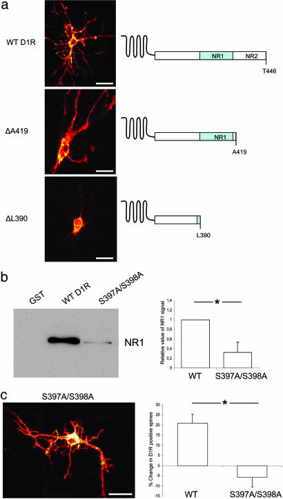
Fig. 5.
Role of D1R C terminus on receptor localization and binding to NR-1. (a) Representative confocal images of neurons transfected with WT D1R, ΔA419, or ΔL390, fused to Venus. Truncation of the C terminus and binding sites for NMDA receptor subunit NR1 (L387–L416) and NR2A (S417–T446) are indicated. (Scale bar: 50 μm.) (b) Western blot of NR1 subunit after GST pull-down from rat striatal lysate using WT GST-D1R-L387-L416 (WT) or -S397A/S398A, or GST alone. The immunoblots were analyzed by using densitometry, and quantitation is shown in the bar graph (*, P < 0.01). (c) Confocal image of striatal organotypic neuron transfected with D1R-S397A/S398A. The bar graph compares the percentage change in D1R-positive spines in response to NMDA treatment for neurons expressing WT D1R (WT) or D1R-S397A/S398A (*, P < 0.001). (Scale bar: 50 μm.)

