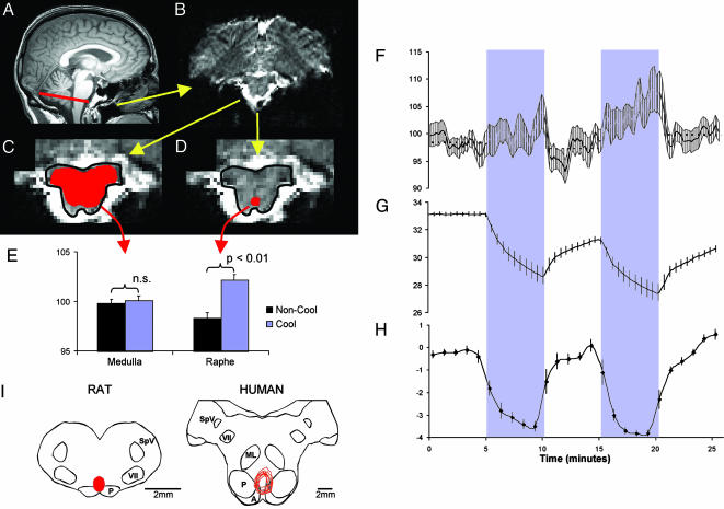
Fig. 1.
Figure showing process for definition of ROIs, responses to skin cooling, and comparative functional anatomy of medullary raphé. (A) Midline sagittal T1-weighted brain image, indicating the rostral medullary slice containing the ROIs (red line). (B) Echoplanar MRI of the rostral medullary slice in the same subject showing the dorsal surface at the top of the image. (C and D) Expanded view of the brainstem from B, highlighting the medullary outline (drawn in black) and the medullary (C) and raphé (D) ROIs drawn in red. (E) Histogram of mean BOLD signal from the medullary and raphé ROIs during cooling (blue bars) and noncooling (black bars) periods in nine subjects. Significant differences are indicated. (F–H) Time profiles of the mean raphé BOLD signal (F), mean skin temperature (G), and mean subjective ratings of skin thermal sensation (H) in nine subjects. The two 5-min cooling periods in the protocol are indicated by a blue stippled background. In all panels, the error bars denote SEM. (I Left) Cross section drawing of rat rostral medulla, indicating the region found to be essential for cold-driven vasoconstriction and thermogenesis [the red area indicates the overlying raphé pallidus nucleus (3, 7)]. (I Right) Corresponding section of human medulla (drawn with reference to Blessing (ref. 47, p. 379) and Duvernoy (ref. 31, pp. 54 and 123) on which are plotted (in red) the nine raphé ROIs analyzed in the present study. A, arcuate nucleus; ML, medial lemnicus; P, pyramidal tract; SpV, spinal trigeminal nucleus; VII, facial nucleus.

