
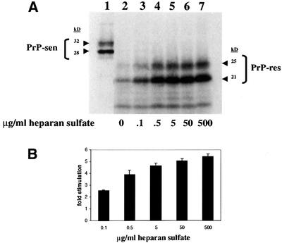
Fig. 1. HS stimulation of hamster GPI(–) PrP cell-free conversion. (A) Effect of increasing concentrations of HS on cell-free conversion. HS was added to conversion reactions at the concentrations indicated (lanes 2–7). An aliquot of the pre-incubation mixture representing 20% of the input [35S]PrP-sen is shown in lane 1. The two bands at ∼28 and ∼32 kDa show unglycosylated and partially glycosylated [35S]PrP-sen, respectively. The radiolabeled protein bands at ∼21 and ∼25 kDa after PK digestion demonstrate that the input [35S]PrP-sen had acquired limited PK resistance similar to brain-derived PrP-res (Kocisko et al., 1994). (B) Fold stimulation of cell-free conversion by increasing concentrations of HS. Error bars show the standard deviation (n = 3).