
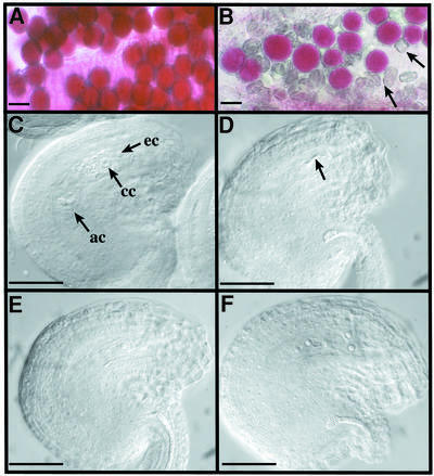
Fig. 2. Male and female gametophyte observation. Viability of mature pollen grains of Ws (A) or spo11-1-1 (B) plants after Alexander staining. Cytoplasm from viable pollen grains is coloured pink. Arrows point to dead pollen grains. The scale bar represents 25 µm in (A) and (B). (C–F) DIC observation of mature ovules of Ws (C) or spo11-1 plants (D–F) after clearing. In a wild-type mature ovule (C), we can observe some of the seven cells of a mature embryo sac (ec, egg cell; cc, central cell; ac, antipodal cells; synergids are out of focus). At the same stage of development, we observe various differentiation stages in the mutant. As examples, in (D) no embryo sac has developed and only degenerated cells can be seen (arrow), (E) shows a one-nucleus embryo sac and (F) shows a four-nuclei embryo sac. For (C–F), the scale bar represents 35 µm.