
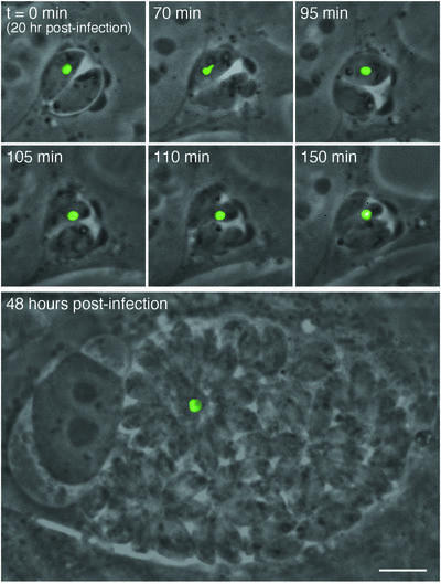
Fig. 3. Unequal segregation of the apicoplast induced by ACP–GFP–mROP1. RH parasites were transiently transfected with ACP–GFP–mROP1 and inoculated into an HFF cell monolayer. Top: beginning 24 h post-transfection/infection (at which point expression of the transgene becomes apparent), parasite division was monitored by time-lapse video microscopy (time points indicated in each frame). Bottom: a different vacuole at 48 h post-transfection, containing ∼64 parasites, only one of which contains an apicoplast. All images are at the same magnification. Bar = 5 µm.