
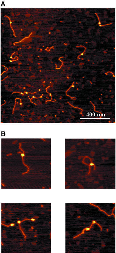
Fig. 7. Atomic force images of UvrA2B complexes formed on the non-damaged DNA substrate. The colour scale ranges from 0.0 to 3.0 nm (from dark to bright). (A) Image of UvrA2B–DNA complexes. (B) Zoomed images of DNA molecules with one or two UvrA2B complexes. Image size, 400 nm.