
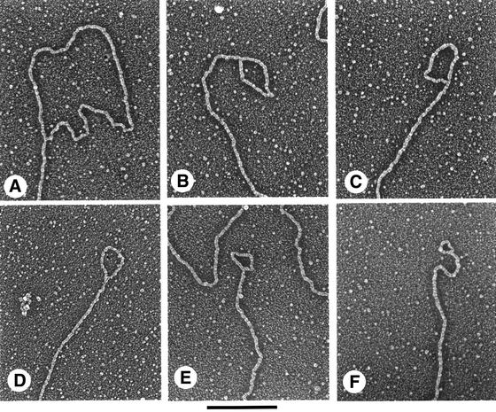
Fig. 2. Visualization of t-loops from T.brucei DNA photo-cross-linked with AMT and UV light. Trypanosomes were permeabilized with digitonin and treated with AMT and UV light, followed by endonuclease cleavage of purified DNA and isolation of the telomeric restriction fragments by gel filtration. DNA fragments were prepared for EM by spreading on a denatured film of cytochrome c protein and rotary shadowcasting with platinum:paladium. Shown in reverse contrast. t-loops shown in (A–F) measured 6.3, 1.75, 1.5, 1.2, 0.99 and 0.63 kb, respectively. Bar is equivalent to 1 kb.