
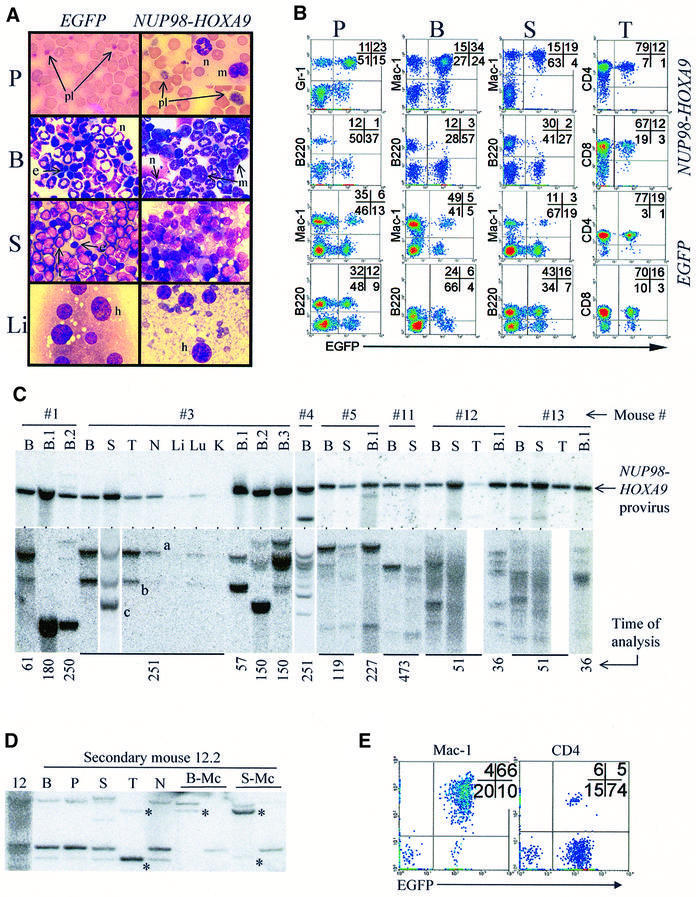
Fig. 2. Characterization of NUP98–HOXA9-induced myeloproliferation in mice. (A) Wright-stained bloodsmear (125×) and cytospin (100×) preparations from representative NUP98–HOXA9 and EGFP control mice. Peripheral blood, P; bone marrow, B; spleen, S; liver, Li; platelets, pl; neutrophils, n; monocytes, m; lymphocytes, l; erythroblasts, e. (B) FACS analyses of hemopoietic organs (peripheral blood, P; bone marrow, B; spleen, S; and thymus, T) of representative NUP98–HOXA9 (#12, see Table I) and EGFP (EGFP-3 group, Table I) control mice, staining for Mac-1, Gr-1, B220, CD4 and CD8. Infected cells are identified by EGFP fluorescence. (C) In NUP98–HOXA9 mice, the provirus is intact (top panel) and the population of NUP98–HOXA9-infected cells is oligoclonal (bottom panel). Top panel: Southern blot analysis assessing the integrity of the provirus shows that the 5.1 kb NUP98–HOXA9-bearing provirus is present and intact in the bone marrow (B) of all mice analyzed, and is also detected in the spleen (S), thymus (T), lymph nodes (N) and lung (Lu), but only weakly so in the liver (Li) and not at all in the kidneys (K). With the exception of one mouse (#4, lower band), no significant amount of rearranged provirus is detected. Bottom panel: Southern blot analysis surveying the integration sites of the proviruses. The time at which the mice were analyzed (in days post-transplantation) is shown at the bottom. For mice 1–4, membranes were hybridized to the Neo probe, and for all other mice the EGFP probe was used. Clones that contribute to myeloid colonies in secondary transplants are totipotent (D and E). (D) Clonal analysis by Southern hybridization of DNA from primary mouse #12 and a secondary recipient (12.2). B, bone marrow; P, peripheral blood; S, spleen; T, thymus; N, lymph nodes; B-Mc and S-Mc, DNA from cells harvested from bone marrow and splenic myeloid colonies grown in methylcellulose cultures, respectively. Asterisks identify totipotent clones by virtue of contribution to myeloid colonies and to reconstitution of the thymus. (E) FACS analysis confirms the contribution of NUP98–HOXA9-expressing (EGFP+) cells to myeloid (Mac-1) and lymphoid (CD4) lineages in the spleen of mouse #12.2. For mouse numbers refer to Table I.