FIG. 2.
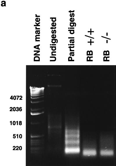
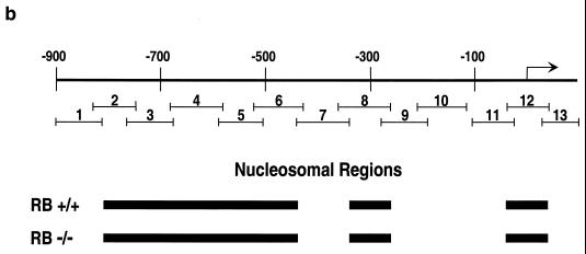
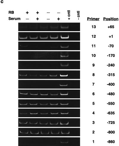
Determination of nucleosome positioning on the mouse cyclin E promoter in RB+/+ and RB−/− MEFs. (a) Micrococcal nuclease digestion of RB+/+ and RB−/− MEF nuclei to produce mononucleosomal DNA. Samples were from formaldehyde-cross-linked chromatin that either was not digested with micrococcal nuclease (undigested) or was subjected to micrococcal nuclease digestion to produce partially digested chromatin (partial digest) or predominantly mononucleosome-size chromatin (RB+/+ and RB−/−). Marker sizes are in base pairs. (b) Illustration of the cyclin E promoter. The amplified region of each PCR product is shown with its corresponding number (see the text). The resulting nucleosomal regions are diagrammed as solid black bars. (c) Results of PCR analyses, with the region of the cyclin E promoter and the number corresponding to the amplified PCR product listed. RB+/+ and RB−/− MEFs were arrested in G0 (−serum) or induced to late G1 (+serum). cntl, control.


