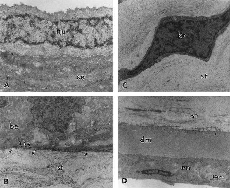
FIG. 6.
Transmission electron microscopy of a transected cornea from a 51-day-old ALDH3a1 knockout mouse showing normal structure. (A) Superficial epithelium consisting mainly of nucleus-free squamous epithelium (se) but occasionally showing a nucleated cell (nu); (B) basal portion of the basal epithelum (be) and adjacent anterior stroma (st) with normal structures for a mouse, including basal lamina (arrowheads) and adjacent hemidesmosomes (arrows), but no Bowman’s membrane; (C) normal keratocyte (kr) in the central stroma (st); (D) normal-appearing posterior stroma (st). Descemet’s membrane (dm), and endothelium (en). The corneas of heterozygote and homozygote wild-type mice were similarly normal in structure. Magnification, ×15,000. Calibration bar, 1.0 μm for all micrographs.

