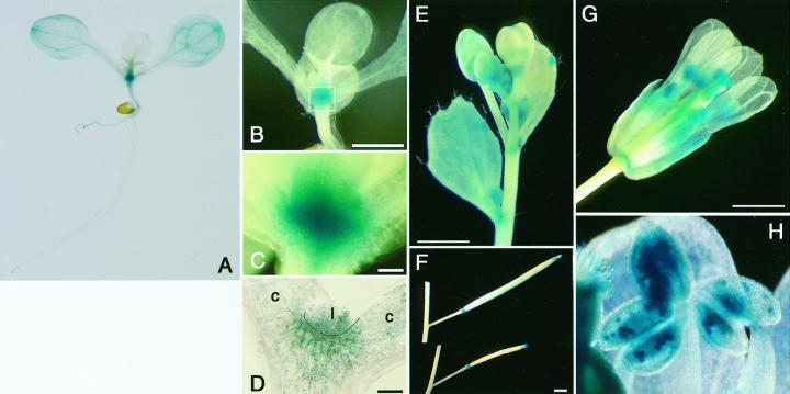
Figure 3.
Histochemical GUS staining of AtMinE1 promoter::uidA-transgenic Arabidopsis plants. GUS staining patterns are shown for whole seedlings (A–C), a section of meristematic region of a seedling (D), and plant organs (E–H); cauline leaf and inflorescence (E), silique (F), open flower (G), and anther at higher magnification (H). C represents an enlargement of the boxed area in B. c, Cotyledon; l, leaf. Scale bars represent 1 mm.

