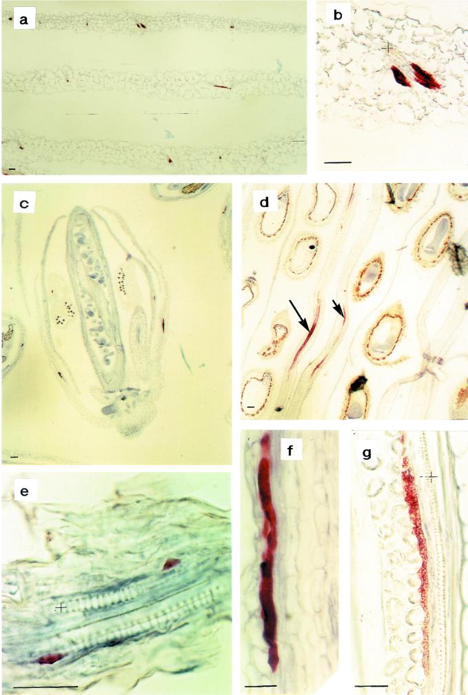
Figure 3.
Immunohistochemical analysis of myrosinase in Arabidopsis using the monoclonal antibody 3D7. In fully expanded leaves from 25-d-old plants, myrosinase were found in often pair-wise-occurring idioblastic cells of the phloem parenchyma (a and b). b, Larger magnification of a. In flower buds, some cells contained myrosinase (c and f), even in the developing petal and sepal (c). In 10-d-old siliques, the myrosinase containing idioblastic phloem parenchyma cells were larger and the staining was more granulated (d and g). d, Arrows indicate long myrosinase-expressing cells. e, The yellowish staining outside the endosperm is retained unoxidized substrate and is therefore regarded as background. In 5-d-old seedlings, developing myrosinase-containing phloem cells were only found in the axis. Xylem is marked with + and the size bars correspond to 10 μm.

